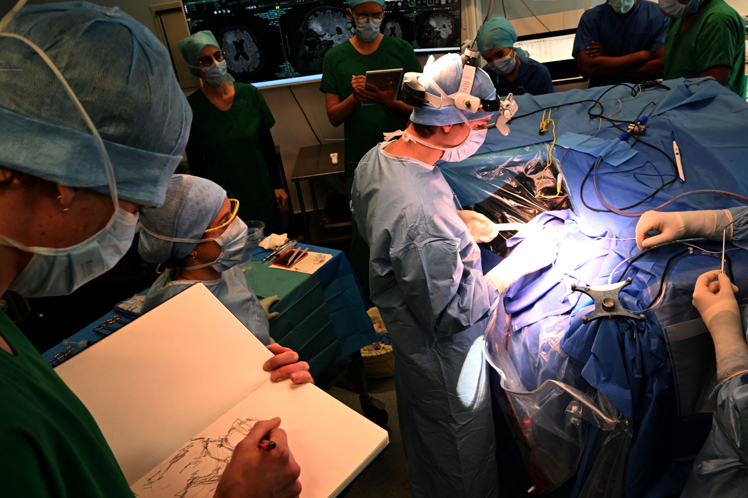
In May 2026, surgeons at London’s National Hospital for Neurology and Neurosurgery performed a first-of-its-kind procedure: they used a real-time computer vision AI system during a live brain operation to remove a pituitary tumor. The patient, Rhys Hibbert, woke up seeing clearly for the first time in over a year. Unlike traditional surgical navigation that relies on pre-operative MRI or CT scans, this AI watched live video from the operating microscope and color-coded critical anatomy in real time.
This marks a major leap from static imaging to dynamic, intraoperative guidance. For surgeries where a millimeter mistake means permanent blindness, the difference is enormous.
What Happened
Rhys Hibbert, 48, discovered he had an 11-millimeter pituitary adenoma after collapsing from a seizure in December 2024. The tumor pressed against his optic chiasm, gradually robbing him of sight over a year. Surgeons needed to remove the tumor without damaging the optic nerves, blood vessels, and chiasm—all packed within a 1-millimeter margin.
The UCL-developed AI system processed live video from the surgical microscope, instantly identifying and highlighting the tumor, nerves, and blood vessels. It overlaid color-coded boundaries directly on the surgeon’s view, updating at video frame rates. This allowed the team to navigate continuously without pausing to match pre-op scans to a deformed surgical field—a common source of error.
The patient recovered fully and reported restored vision immediately after surgery. The case was published as a world-first for real-time computer vision in neurosurgery.
My Take
This is the kind of AI application that actually saves lives—no hype, no API calls for chat. It’s a narrow, well-trained vision model solving a specific, high-stakes problem. The key insight is moving from “look at this scan” to “look at what’s on the screen right now.” Brains shift during surgery. Pre-op scans are approximations. Real-time video is truth.
For developers and engineers, this signals where edge AI is heading: embedded, low-latency vision systems running in surgical microscopes, not cloud servers. The inference has to happen locally, at 30+ fps, with absolute reliability. That’s a hard engineering problem, and UCL appears to have cracked it.
What to Watch
- Real-time surgical AI becomes a standard tool for any operation involving critical nerves or vessels, not just pituitary surgeries.
- Expect a wave of startups building specialized vision models for different surgical disciplines—orthopedics, ophthalmology, neurosurgery.
- Regulatory bodies will need to update approval frameworks for AI that makes live, intraoperative decisions rather than pre-op analysis.